Vertebrate brain theory
for the European Union’s Human Brain Project
ISBN 978-3-00-064888-5
4.6 The dorsal ventricular crest of reptiles and birds - a convergence system and a vertical convergence grid
Similar to the frontal cortex, a flat convergence system also formed in reptiles and birds for further processing of the output of the thalamic nucleus ventralis lateralis - which received its signals from the spinocerebellum. However, there was a serious topological difference.
We consider the initial situation before signal divergence in the nucleus olivaris and restrict ourselves to the muscle spindle signals. For a simple joint, there were two muscles working against each other to adjust the joint angle.
These signals formed a bivalent signal pair per joint, which reached the spinocerebellum via two climbing fibres. From this pair of signals, the nucleus olivaris and thus also the cerebellum produced a whole set of signals through signal divergence. And exactly this set of signals had to be reduced again to the two output signals in the convergence circuit. Mammals realized the convergence circuit in the frontal cortex. Reptiles and birds developed other procedures.
For this purpose, the signals of this signal set were sent headlong in the secondary turning loop and transferred to a neuron set in which the neurons were arranged in a vertical line one above the other. The neighborhood of the signals was preserved as it was in the nucleus olivaris or in the cerebellar nucleus. Exactly above and below these input neurons there was one output neuron each, one above and one below. And all input neurons of the signal set were excitedly connected to both output neurons. So all vertically arranged input neurons were connected to the two corresponding output neurons of the bivalent signal pair. Thus, the cerebellar signal divergence was reversed.
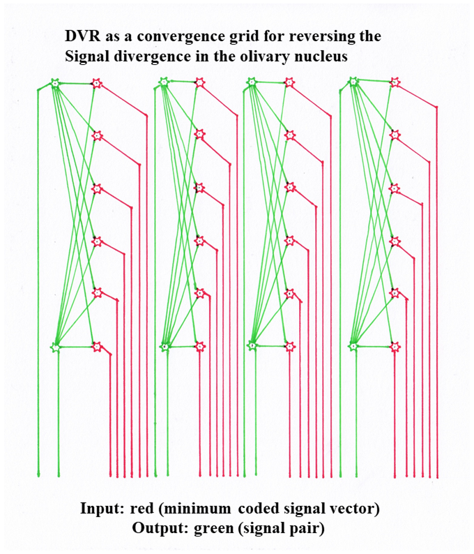
Figure 42 - DVR as Convergence Grid
This convergence structure was located inside the neural tube, where all neurons were generally located, while their axons were located outside. Of course, it also took up a certain amount of space. The further the signal divergence in the nucleus olivaris progressed and the more input axons were present, the thicker this neuron layer of input neurons inside the layer and output neurons on the outer surfaces became. The structure took up more and more space and therefore grew into the free ventricular space that filled the interior of the hollow neural tube. It was located somewhat dorsally. Therefore, this convergence system was later called the dorsal ventricular crest (DVR). He realized the solution to the convergence problem for the thalamic output of reptiles and birds, which originated from the cerebellum.
Since the DVR is also a non-markless structure, because it must have distance-dependent attenuation according to the cable equation for non-markless fibers, there is of course a problem for the rather long input and output axon lines. These are non-markless, but must not have a high attenuation. For this reason the input and output axons are quite thick and therefore transport the action potentials over longer distances. They are clearly visible in the microscopic picture as rather thick axons of different lengths running parallel to each other.
Theorem of the DVR as convergence lattice
The dorsal ventricular crest (DVR) of reptiles and birds is a vertical convergence grid, which corresponds to the frontal cortexof mammals in its function, but is not oriented horizontally in the plane, but vertically in space. It reverses the signal divergence of the nucleus olivaris so that the output can be used for motor control.
Since the different modalities have not yet been separated in the neural tube, forming modality layers there, the DPR consists of several modality layers stacked on top of each other, each of which has the appearance shown in the above diagram. Due to the large thickness of this structure, the incoming and outgoing axons must be quite thick to prevent too much attenuation. They form prominent objects in the microscopic image, while the actual convergence layers have a rather homogeneous appearance.
Monograph of Dr. rer. nat. Andreas Heinrich Malczan